Category:Cell biology
Перейти до навігації
Перейти до пошуку

| Category Cell biology on sister projects: | |||||||||
|---|---|---|---|---|---|---|---|---|---|
Wiktionary |
Commons | ||||||||
розділ біології  | |||||
| Завантажити медіафайл | |||||
| Вимова (аудіофайл) | |||||
|---|---|---|---|---|---|
| Є одним із | |||||
| Є підкласом | |||||
| Частина від | |||||
| Не плутати з | |||||
| |||||
Підкатегорії
Показано 118 підкатегорій із 118.
*
A
- A549 cell line (2 F)
- Cell aggregation (103 F)
- Cell aging (17 F)
- Anisotropy in biology (8 F)
B
- Biological metamorphosis (23 F)
- Brochosome (6 F)
C
- Cell count (69 F)
- Cell degranulation (15 F)
- Cell disruption (17 F)
- Cell dynamics (5 F)
- Cell enlargement (19 F)
- Cell extracts (6 F)
- Cell fate determination (154 F)
- Cell fusion (109 F)
- Cell potency (52 F)
- Cell tracking (43 F)
- Cellular immunity (8 F)
- Cellular stress responses (247 F)
- Chemiosmosis (18 F)
- Colony-forming units assays (21 F)
- Cytolysis (3 F)
D
E
- Endomembrane system (10 F)
- Extracellular traps (22 F)
F
G
- Goodsell molecular landscape (14 F)
H
I
- Intracellular space (82 F)
M
- Media from BMC Cell Biology (220 F)
- Media from Cell & Bioscience (4 F)
- Media from Cell & Chromosome (4 F)
- Media from Cell Regeneration (2 F)
O
- Oogenesis (42 F)
- Osmotic shock (6 F)
P
- Cell-penetrating peptides (12 F)
- Phytoliths (14 F)
- Plasmolysis (24 F)
- Prebiotic (316 F)
- Protoplasts (18 F)
R
S
- Cell shape (259 F)
- Single-cell analysis (80 F)
- Subcellular fractions (57 F)
- Subcellular localization (10 F)
- Surface properties (biology) (7 F)
- Cell survival (228 F)
T
V
- Videos of cell biomechanics (13 F)
Z
- ZTZ der Uniklinik Freiburg (6 F)
Файли в категорії «Cell biology»
Показано 200 файлів цієї категорії (із 540).
(попередня сторінка) (наступна сторінка)-
 022S.jpg 1772 × 1772; 3,11 МБ
-
 023S.jpg 1772 × 1792; 612 КБ
-
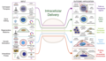 1 - ID applications.png 1662 × 950; 459 КБ
-
 200801large.jpg 600 × 457; 298 КБ
-

-
 208031 EPFL David Suter Sox2.jpg 1304 × 734; 52 КБ
-
 3eme vague.png 550 × 357; 7 КБ
-
 41598 2018 20107 Fig3 HTML.jpg 660 × 315; 167 КБ
-
 46085 orig.jpg 2816 × 2112; 412 КБ
-
 72hr sodyum48 saat.jpg 1600 × 1200; 272 КБ
-
 A Critical-like Collective State Leads to Long-range Cell Communication in Dictyostelium discoideum Aggregation.pdf 1275 × 1650, 19 сторінок; 3,34 МБ
-
 A sol mit Paramecium.jpg 1327 × 885; 241 КБ
-
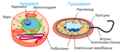
 A) Endocitosis y b) exocitosis.jpg 818 × 529; 109 КБ
-
-
 A549 bridges -- 7-28-at1622.jpg 1392 × 1040; 160 КБ
-
 A549 bridges -- 7-28-at1623.jpg 1392 × 1040; 149 КБ
-

-
 Adrenergic signaling on natriuretic peptides.jpg 2958 × 2165; 711 КБ
-
 Alcatel One Touch 720.pdf 1239 × 1752, 4 сторінки; 48 КБ
-
 Allosteric regulation mode, feedback inhibition and its reversal.png 709 × 499; 72 КБ
-
 ALPSc.jpg 373 × 197; 19 КБ
-
 Alveolar sac region of the lung - TEM.jpg 1560 × 1257; 522 КБ
-
 Amiloplastos de células de papa.jpg 302 × 325; 53 КБ
-
 Animal Cell Structure.png 724 × 484; 145 КБ
-
 Annotated structure of eRF1.jpg 600 × 361; 81 КБ
-
 Annular Gap Junction Vesicle.jpg 205 × 153; 61 КБ
-
 Anthracologie-exemple.jpg 860 × 860; 848 КБ
-
 Anthracologie-exemple2.jpg 896 × 896; 786 КБ
-
 Anticossos anticardiolipina.jpg 427 × 286; 61 КБ
-
 Apical Constriction.jpg 242 × 300; 80 КБ
-
 Apicalconstriction fig1.jpg 417 × 467; 82 КБ
-
 Apicalconstriction fig2.jpg 393 × 437; 82 КБ
-
 Apoptotic DNA Laddering.png 178 × 193; 20 КБ
-
 ART SCIENCE Craig Hilton 'The Immortalisation of Billy Apple' 01.jpg 3264 × 2448; 2,91 МБ
-
 ART SCIENCE Craig Hilton 'The Immortalisation of Billy Apple' 02.jpg 3264 × 2448; 3,01 МБ
-
-
 Aurora B localization.jpg 93 × 349; 8 КБ
-
 Autophagy and apoptosis.png 678 × 339; 97 КБ
-
 Autophagy and cancer.jpg 985 × 380; 76 КБ
-
 Autophagy in plants.jpg 600 × 424; 53 КБ
-
 Autophagy's function.gif 200 × 98; 11 КБ
-
 Autophagy.png 2012 × 1636; 1,59 МБ
-
 Axopodium Mikrotubuli.jpg 748 × 682; 269 КБ
-
 Banner cell biology.png 1003 × 100; 45 КБ
-
 BarrBodyBMC Biology2-21-Fig1.jpg 1200 × 1417; 298 КБ
-
 Betareg.PNG 523 × 511; 18 КБ
-
 BiggeggSH-SY5Y.jpg 1600 × 1200; 426 КБ
-
 BioArchive.jpg 263 × 342; 31 КБ
-
 Blood cell crossing vascular sinus wall - TEM.jpg 1560 × 1254; 518 КБ
-
 Boveri-signature.jpg 424 × 91; 20 КБ
-
 Branching morphogenesis in 3D cell culture.jpg 778 × 434; 98 КБ
-
 Brefeldin A Inhibition of Intracellular Vesicle Transport.png 591 × 271; 23 КБ
-
 Brochosome model1.jpg 729 × 360; 40 КБ
-
 BrUpolIIc.jpg 406 × 233; 33 КБ
-
 BS-Fig1.jpg 658 × 336; 105 КБ
-
 Cajal frontal lobe.gif 250 × 442; 39 КБ
-
 Cajal Purkinje.gif 300 × 332; 32 КБ
-
 Calcium Signaling Pathway.png 1526 × 907; 180 КБ
-
 Cancer type vs frequency.png 943 × 354; 25 КБ
-
 Catenin-humanendothel.jpg 800 × 634; 376 КБ
-
 CBDS.Mirmira-300x167.png 300 × 167; 58 КБ
-
 Cel cible compétente.png 854 × 165; 6 КБ
-
 Celcultuuroven.JPG 2560 × 1920; 1,94 МБ
-
 Celkweekverversing.jpg 1920 × 2560; 279 КБ
-
 Cell (PSF).jpg 969 × 430; 386 КБ
-
 Cell 1 a.jpg 373 × 200; 46 КБ
-
 Cell 1.jpg 1500 × 805; 654 КБ
-
 Cell structure.jpg 1280 × 720; 127 КБ
-
 Cell-organelles-labeled.png 1050 × 1024; 743 КБ
-
 Cell-organelles.webp 600 × 375; 15 КБ
-
 Cell-shape-mitosis.png 633 × 668; 167 КБ
-
 Cell-type specificity of TIP-YFP expression in the root axis cropped.jpg 1050 × 565; 120 КБ
-
 Cell-type specificity of TIP-YFP expression in the root axis.jpg 1200 × 2007; 554 КБ
-
 Cell-universe.jpg 1398 × 1045; 51 КБ
-
 Cells detention centers 2.jpg 527 × 265; 87 КБ
-
 Cells detention centers 3.jpg 300 × 151; 65 КБ
-
 Cells detention centers.jpg 344 × 172; 59 КБ
-
 Cells in space.JPG 1280 × 960; 398 КБ
-
 Cellsize it.jpg 310 × 199; 10 КБ
-
 Cellsize PL.png 310 × 199; 72 КБ
-
 Cellsize.jpg 310 × 199; 51 КБ
-
 Celltype zh.png 1280 × 535; 176 КБ
-
 Celltypes rus.png 468 × 202; 51 КБ
-
 Cellular Dewetting.jpg 606 × 102; 14 КБ
-
 Cellular Structure (2).jpg 5312 × 2988; 4,08 МБ
-
 Cellular Structure.jpg 5243 × 2854; 3,11 МБ
-
 Cellular tight junction-cz.svg 499 × 646; 145 КБ
-
 CELLULES ROUGES.jpg 785 × 785; 466 КБ
-
 Cellules souches embryonnaires HD90.jpg 2048 × 1536; 623 КБ
-
 Cellules souches embryonnaires HD90.tif 2048 × 1536; 6 МБ
-
 Celulasok.jpg 2491 × 1072; 3,59 МБ
-
 Chaperiony.png 794 × 1123; 77 КБ
-
 CHIB.-Sander-300x226.jpg 300 × 226; 25 КБ
-
 CHIB.-Stabler.jpg 1362 × 627; 675 КБ
-
 Chloride cell.jpg 574 × 480; 276 КБ
-
 Cho cells adherend1.jpg 1280 × 960; 387 КБ
-
 Cho cells adherend2.jpg 1280 × 960; 240 КБ
-
 Chromaffin cell imaged with DIC and IRM.png 330 × 212; 63 КБ
-
 Cineálacha éagsúla ceall.jpg 450 × 188; 43 КБ
-
 Città della Scienza Catania 2.jpg 2896 × 1944; 1,49 МБ
-
 Cleavage-furrow.JPG 449 × 447; 23 КБ
-
 Clonal expansion and monoclonal versus polyclonal proliferation.PNG 660 × 510; 34 КБ
-
 Cluster of Solenocytes.jpg 862 × 930; 152 КБ
-
 CnGRASP55domainsc.jpg 763 × 473; 112 КБ
-
 CollagenMegaCarrierc.jpg 553 × 249; 45 КБ
-
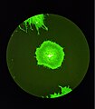
 Colonies of Madin-Darby Canine Kidney cells grown in tissue culture.jpg 1030 × 1030; 263 КБ
-
 Color fungi.jpg 1503 × 1127; 452 КБ
-
 Comparison chrx.jpg 1114 × 729; 491 КБ
-

-
 Complete Hydatidiform Mole (38711526030).jpg 1732 × 2048; 1,34 МБ
-
 Conger type callus 3ms White Light.TIF 2048 × 1536; 9,01 МБ
-
 Conidiospore-hyaloperonospora-parasitica-appressorium.jpg 590 × 233; 53 КБ
-
 Cotyledon-Cercis siliquastrum.jpg 772 × 516; 243 КБ
-
 Coxiella burnetii, the bacteria that causes Q Fever.jpg 2424 × 2032; 1,19 МБ
-
 CPE rounding.jpg 1200 × 900; 366 КБ
-
 CPE syncytium.jpg 1200 × 900; 468 КБ
-
 Cresta Localiza ATPsintasa.png 580 × 440; 126 КБ
-
 Cresta Mitocondrial.png 1123 × 794; 293 КБ
-
 Crosssectionroottooth11-24-05.jpg 1263 × 1053; 259 КБ
-
 Crosstalk TM.png 1588 × 616; 182 КБ
-
 Cryptosporidium parvum auramine-rhodamine labeled.jpg 300 × 308; 6 КБ
-
 CTAR Powers.png 1173 × 639; 1,19 МБ
-
 CTAR.-Herrera-300x165.png 300 × 165; 50 КБ
-
 Cyborg Cell characteristics.jpg 501 × 510; 143 КБ
-
 Cytogenetic Telomere Arrays (2005).jpg 990 × 1333; 200 КБ
-
 Cytokinesis-electron-micrograph.jpg 745 × 451; 200 КБ
-
 Cytoneme.tif 1400 × 796; 1,28 МБ
-
 Células en un tejido normal.jpg 1280 × 720; 39 КБ
-
 Dedication to Plant Cell Biology Second Edition, Elsevier 2019.jpg 4383 × 6284; 3,32 МБ
-
 Dental Pulp cultured by MLM (3D vs. 2D) - 5 Days.tiff 1353 × 1112; 1,18 МБ
-
 Desmosome cell junction cs.svg 556 × 588; 124 КБ
-
 Desplazamiento del punto central de la célula.gif 724 × 427; 11 КБ
-

-

-
 Different ways mtDNA moves into the nucleus.PNG 1280 × 720; 89 КБ
-
 Differentiation of Stem Cells Into Neurons.jpg 1884 × 835; 845 КБ
-
 Differentiation.jpg 5454 × 3225; 31,2 МБ
-
 DnTRFc.jpg 462 × 259; 34 КБ
-
 Domain architecture and structure of C-terminal EHD proteins.gif 583 × 522; 41 КБ
-
 Drachenbaum Bewurzelung-in-Wasser Kallus SL273382.JPG 2304 × 3072; 2,23 МБ
-
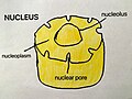 Drawing of nucleus.jpg 2448 × 1836; 1,05 МБ
-
 Duplicazione dei plasmidi.png 800 × 700; 223 КБ
-
 Débit de Filtration Glomérulaire Inkscape.png 2500 × 1500; 544 КБ
-
 Débit de Filtration Glomérulaire librsvg.png 2500 × 1500; 458 КБ
-
 Débit de Filtration Glomérulaire rendersvg.png 2500 × 1500; 476 КБ
-
 EHD proposed mechanism.jpg 1280 × 373; 70 КБ
-
 Einzeller 800 fach Streiflicht.jpg 1952 × 1372; 409 КБ
-
 Ekstratsellulaarne maatriks.png 800 × 554; 97 КБ
-
 Electron micrograph of a Fractone.jpg 3662 × 3472; 2,37 МБ
-
 Embolized Uterus (44319080955).jpg 1270 × 1124; 808 КБ
-
 Embolized Uterus (44319081065).jpg 1352 × 1124; 811 КБ
-
 Embolized Uterus (44507244194).jpg 1124 × 1208; 885 КБ
-
 EndoERGICNIHlc.jpg 273 × 291; 23 КБ
-
 Endometrial Intraepithelial Neoplasia (EIN) (46262991251).jpg 2347 × 1706; 1,61 МБ
-
 Endosymbiose.jpg 1899 × 285; 198 КБ
-
 Entropy in living cells.jpg 897 × 606; 93 КБ
-
 Eosonophilic promyelocyte.png 115 × 104; 12 КБ
-
 Epitelo-mezenchymální tranzice.gif 822 × 214; 36 КБ
-
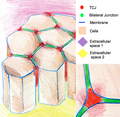 Epithelium TCJ.png 755 × 736; 969 КБ
-
 Epithelzelle 43729.jpg 531 × 384; 24 КБ
-
 Epithelzellen16631.jpg 531 × 384; 33 КБ
-
 ER-containing autophagosome.png 1417 × 1770; 2,06 МБ
-
 ERK propagation waves from the wound edge.png 1340 × 547; 1,07 МБ
-
 Ethanol-Bilayer.png 1194 × 639; 265 КБ
-

-
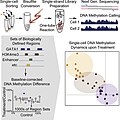 Farlik abstract.jpg 375 × 375; 71 КБ
-
 FGF Pathway tm.png 1242 × 698; 58 КБ
-
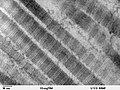 Fibers of Collagen Type I - TEM .jpg 640 × 480; 105 КБ
-
 Fibers of Collagen Type I - TEM.jpg 640 × 480; 100 КБ
-
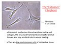 Fibroblast-2.jpg 2999 × 2249; 693 КБ
-
 Flow cytometer structure.png 865 × 599; 1,98 МБ
-
 Flowcytometer MoFlo (DAKO cytomation).jpg 320 × 426; 21 КБ
-
 Flower petal cells image 2.jpg 3072 × 4096; 5,6 МБ
-
 Flower petal cells.jpg 3072 × 4096; 4,75 МБ
-
 Fluorescence.microscope1.jpg 2226 × 2544; 1,09 МБ
-
 Fluorescence.microscope2.jpg 2428 × 2475; 1,21 МБ
-
 Fluorescence.microscope3.jpg 2436 × 2393; 1,26 МБ
-
 Food particle from a colonoscopy specimen (34016474661).jpg 1480 × 811; 369 КБ
-
 Foreign Body Granuloma on the Peritoneum (41962302110).jpg 2402 × 1458; 1,28 МБ
-
 Formacao Mesossomo.png 900 × 800; 63 КБ
-
 Formation of the new blood vessels.jpg 1920 × 1200; 2,81 МБ
-
 FTIR of cell.png 3025 × 2266; 2,28 МБ
-
 Functions of DS.PNG 585 × 402; 104 КБ
-
 Fusion secuencia.jpg 2084 × 1828; 2,62 МБ
-
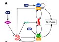 G1-S cell cycle regulation.jpg 800 × 576; 68 КБ
-
 G2-M Bistability.png 1347 × 1012; 49 КБ
-
 Ganglion Cyst of Hand (35295313226).jpg 2102 × 1669; 659 КБ
-
 Gauchdisease.jpg 314 × 209; 74 КБ
-
 GemIdentComposite.jpg 744 × 683; 125 КБ
-
 Gephyrinc.jpg 542 × 347; 52 КБ
-
 Gewebetypen des Blattes.jpg 3508 × 2480; 1,28 МБ
-
 Glycophosphatidylinositol anchor.tif 585 × 432, 2 сторінки; 514 КБ
-
 GMAP210ALPSc.jpg 376 × 188; 19 КБ
-
 GMAP210c.jpg 420 × 71; 11 КБ
-
 Gnf-segmented-41-closeup.png 351 × 236; 83 КБ
-
 GolgiCisConnc.jpg 330 × 123; 21 КБ
-
 GolgiCOGsc.jpg 748 × 324; 84 КБ
-
 GolgiRibbonc.jpg 250 × 143; 23 КБ
-
 GolgiScyl1c.jpg 471 × 296; 46 КБ
-
 GolgiTethersc.jpg 390 × 233; 28 КБ


_Endocitosis_y_b)_exocitosis.jpg)


.jpg)


.jpg)


.jpg)


.jpg)


.jpg)
.jpg)
.jpg)

_(46262991251).jpg)


.jpg)


.jpg)
.jpg)


.jpg)


