File:Renal corpuscle.svg

File originale (file in formato SVG, dimensioni nominali 1 030 × 760 pixel, dimensione del file: 535 KB)
Didascalie
Didascalie
|


|
Questa immagine è stata valutata secondo i criteri per le immagini di valore ed è considerata l'immagine di maggior valore su Commons per l'argomento: Diagram of renal corpuscle. Puoi vedere la sua valutazione qui. |
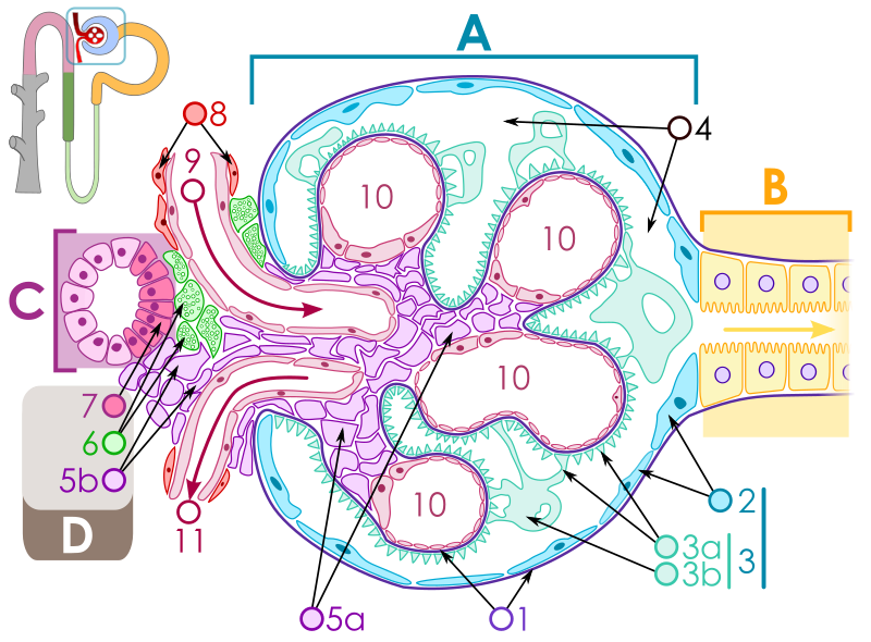
Questa immagine è stata selezionata come Immagine del giorno in data 8 maggio 2009. La didascalia era la seguente: Italiano: Struttura di un corpuscolo renale. Altre lingue:
Afrikaans: Diagram van 'n nierliggaampie Bosanski: Dijagram strukture bubrežnog tjelašca. Čeština: Schéma ledvinového tělíska Deutsch: Schematischer Aufbau des Nierenkörperchens English: Diagram of renal corpuscle structure Español: Diagrama de la estructura del corpúsculo renal Français : Schéma d'un glomérule rénal. Italiano: Struttura di un corpuscolo renale. Magyar: A vesetestecske (Corpusculum renale) felépítésének sematikus ábrázolása Nederlands: schematisch overzicht van het lichaampje van Malpighi Polski: Schemat budowy ciałka nerkowego Português: Diagrama da estrutura de um Corpúsculo de Malpighi. Română: Diagramă a unui nefron. Suomi: Kaavakuva munuaiskeräsen rakenteesta. Македонски: Приказ на составот на едно бубрежно телце Русский: Схема строения почечного тельца. 한국어: 신소체의 구조에 관한 다이어그램. 日本語: 腎小体 構造図 中文: 肾小球结构示意图 中文(繁體): 腎小球結構示意圖 |
Dettagli[modifica]
| Descrizione |
Čeština: Schéma struktury ledvinného tělíska:
English: Diagram of renal corpuscle structure:
Deutsch: Schematischer Aufbau des Nierenkörperchens
Español: Esquema de la estructura del glomérulo renal:
Français : Diagramme structural d'un glomérule rénal:
Italiano: Diagramma strutturale del Corpuscolo renale
Polski: Schemat budowy ciałka nerkowego
Português: Esquema da estrutura do glomérulo renal:
Русский: Схема строения почечного тельца
Українська: Схема будови ниркового тільця
|
||||||||||||||||||||
| Data |
9 novembre 2008 18 gennaio 2009 (update) |
||||||||||||||||||||
| Fonte | Opera propria | ||||||||||||||||||||
| Autore |
M.Komorniczak (polish Wikipedist)
|
||||||||||||||||||||
| Licenza (Riusare questo file) |
Io, detentore del copyright su quest'opera, dichiaro di pubblicarla con la seguente licenza: Questo file è disponibile in base alla licenza Creative Commons Attribuzione-Condividi allo stesso modo 3.0 Unported
|
||||||||||||||||||||
| Altre versioni |
Opere derivate da questo file:
[] SVG[modifica]
PNG[modifica] |
||||||||||||||||||||
| SVG sviluppo | Il codice sorgente di questo file SVG non è valido a causa di I errori. Questo diagramma in grafica vettoriale non valido secondo il W3C è stato creato con Inkscape, o con qualche altro programma.
|
||||||||||||||||||||


- ↑ (2019). "Thick Ascending Limb Sodium Transport in the Pathogenesis of Hypertension". Physiological Reviews 99 (1): 235–309. DOI:10.1152/physrev.00055.2017. PMID 30354966. PMC: 6335098.
- ↑ Tubuloglomerular Feedback - an overview | ScienceDirect Topics.
- ↑ (2014). "Thick Ascending Limb of the Loop of Henle". Clinical Journal of the American Society of Nephrology 9 (11): 1974–1986. DOI:10.2215/CJN.04480413. PMID 25318757. PMC: 4220766.
Cronologia del file
Fare clic su un gruppo data/ora per vedere il file come si presentava nel momento indicato.
| Data/Ora | Miniatura | Dimensioni | Utente | Commento | |
|---|---|---|---|---|---|
| attuale | 21:28, 18 gen 2009 | | 1 030 × 760 (535 KB) | M.Komorniczak (discussione | contributi) | {{Information |Description= |Source= |Date= |Author= |Permission= |other_versions= }} |
| 19:14, 18 gen 2009 |  | 1 030 × 760 (533 KB) | M.Komorniczak (discussione | contributi) | == Opis == {{Information |Description={{pl|1=Schemat budowy ciałka nerkowego <table><tr><td align=left> A - Ciałko nerkowe<br> B - Kanalik proksymalny (kanalik główny)<br> C - Kanalik dystalny (wstawka)<br> D - Aparat przykłę | |
| 18:34, 16 gen 2009 |  | 1 030 × 760 (361 KB) | Eusebius (discussione | contributi) | watermark removed Commons:Watermarks | |
| 00:51, 4 gen 2009 |  | 1 030 × 760 (390 KB) | M.Komorniczak (discussione | contributi) | == Opis|Information == {{Information |Description={{pl|1=Schemat budowy ciałka nerkowego A - Ciałko nerkowe B - Kanalik proksymalny (kanalik główny) C - Kanalik dystalny (wstawka) D - Aparat przykłębuszkowy 1. Błona po | |
| 23:05, 10 nov 2008 |  | 665 × 515 (411 KB) | M.Komorniczak (discussione | contributi) | {{Information |Description={{pl|1=Schemat budowy ciałka nerkowego A - Ciałko nerkowe B - Kanalik proksymalny (kanalik główny) C - Kanalik dystalny (wstawka) 1. Błona podstawna 2. Część trzewna torebki Bowmana 3. Przestrze� |
Impossibile sovrascrivere questo file.
Utilizzo del file
Le seguenti 85 pagine usano questo file:
- Kidney
- Wikimedia logo mosaic
- User:Ash Crow/test
- User:Bugboy52.4
- User:JoKalliauer/SVG test suites/Featured details
- User:JoKalliauer/SVG test suites/resvg Issues details
- User:Lar/Mosaic2
- User:M.Komorniczak
- User:Miya/POTY/2009/FA January
- User:OhanaUnited/Wikispecies logo mosaic
- User:Prolineserver/mosaic
- User:Przykuta/Gender mosaic
- User:Rocket000/SVGs/Biology/Human anatomy
- User:ScotXW
- User:Slaunger/Sandbox/POTY gallery
- User:Twice25/Mosaic
- User talk:Albertus teolog/Archiwum1
- User talk:Eusebius/Archives/2009
- User talk:Eusebius/Archives/Promotions/April 2009
- User talk:M.Komorniczak
- User talk:Symode09/Large
- Commons:Featured picture candidates/File:Renal corpuscle.svg
- Commons:Featured picture candidates/Log/January 2009
- Commons:Featured pictures/Non-photographic media/Computer-generated
- Commons:Featured pictures/chronological/2009-A
- Commons:Picture of the Year/2009/Galleries/2009-A
- Commons:Picture of the Year/2009/Galleries/All
- Commons:Picture of the Year/2009/Galleries/Diagrams
- Commons:Picture of the Year/2009/Galleries/Index/1
- Commons:Picture of the Year/2009/Galleries/Index/Diagrams
- Commons:Picture of the Year/2009/Galleries/Table/200901
- Commons:Picture of the Year/2009/Preparation
- Commons:Picture of the Year/2009/R1/File:Renal corpuscle.svg
- Commons:Picture of the Year/2009/Results/R1/ALL/Table
- Commons:Picture of the Year/2009/Results/R1/Diagrams
- Commons:Picture of the Year/2009/Results/R1/Diagrams/Table
- Commons:Potd/2009-05 (da)
- Commons:Potd/2009-05 (de)
- Commons:Potd/2009-05 (eo)
- Commons:Potd/2009-05 (nl)
- Commons:Potd/2009-05 (pt)
- Commons:Potd/2009-05 (tr)
- Commons:Quality images/Subject/Non photographic media
- Commons:Quality images candidates/Archives January 2009
- Commons:STOP!!!! DO NOT DELETE THIS IMAGE TILL YOU REPLACE IT IN THE WIKIMEDIA LOGO MOSAIC
- Commons:Valued image candidates/IDsec.svg
- Commons:Valued image candidates/Renal corpuscle.svg
- Commons:Valued images by topic/Science/Medicine, biology and biotechnology
- File:CEE CUP 2017.svg
- File:Cialko nerkowe.svg
- File:Corpuscule rénal.svg
- File:Minimal Change Disease Pathology Diagram.svg
- File:Renal corpuscle-en.svg
- File:Renal corpuscle.svg
- File:Renal corpuscle Inkscape.png
- File:Renal corpuscle batik.png
- File:Renal corpuscle librsvg.png
- File:Renal corpuscle rendersvg.png
- Template:Other versions/Renal corpuscle
- Template:Potd/2009-05
- Template:Potd/2009-05-08
- Template:Potd/2009-05-08 (af)
- Template:Potd/2009-05-08 (bs)
- Template:Potd/2009-05-08 (cs)
- Template:Potd/2009-05-08 (da)
- Template:Potd/2009-05-08 (de)
- Template:Potd/2009-05-08 (en)
- Template:Potd/2009-05-08 (es)
- Template:Potd/2009-05-08 (fi)
- Template:Potd/2009-05-08 (fr)
- Template:Potd/2009-05-08 (hu)
- Template:Potd/2009-05-08 (it)
- Template:Potd/2009-05-08 (ja)
- Template:Potd/2009-05-08 (ko)
- Template:Potd/2009-05-08 (mk)
- Template:Potd/2009-05-08 (nl)
- Template:Potd/2009-05-08 (pam)
- Template:Potd/2009-05-08 (pl)
- Template:Potd/2009-05-08 (pt)
- Template:Potd/2009-05-08 (ro)
- Template:Potd/2009-05-08 (ru)
- Template:Potd/2009-05-08 (zh-hans)
- Template:Potd/2009-05-08 (zh-hant)
- Template:Potd/2009-05 (ro)
- Template:Potd/2009-05 (zh-hans)
Utilizzo globale del file
Anche i seguenti wiki usano questo file:
- Usato nelle seguenti pagine di ar.wikipedia.org:
- Usato nelle seguenti pagine di bg.wikipedia.org:
- Usato nelle seguenti pagine di bn.wikipedia.org:
- Usato nelle seguenti pagine di bs.wikipedia.org:
- Usato nelle seguenti pagine di ca.wikipedia.org:
- Usato nelle seguenti pagine di crh.wikipedia.org:
- Usato nelle seguenti pagine di cs.wikipedia.org:
- Usato nelle seguenti pagine di cv.wikipedia.org:
- Usato nelle seguenti pagine di de.wikipedia.org:
- Usato nelle seguenti pagine di de.wiktionary.org:
- Usato nelle seguenti pagine di dv.wikipedia.org:
- Usato nelle seguenti pagine di en.wikipedia.org:
- Wikipedia:Featured picture candidates/Renal Corpuscle
- Wikipedia:Featured picture candidates/February-2009
- User:Daniel Mietchen/Science communication gallery
- User:IONTRANSP
- Wikipedia:WikiProject Anatomy/Resources
- Wikipedia talk:WikiProject Anatomy/Archive 9
- User:Scope creep/userboxes
- User talk:Rhododendrites/Reconsidering FPC on the English Wikipedia
- Usato nelle seguenti pagine di es.wikipedia.org:
Visualizza l'utilizzo globale di questo file.